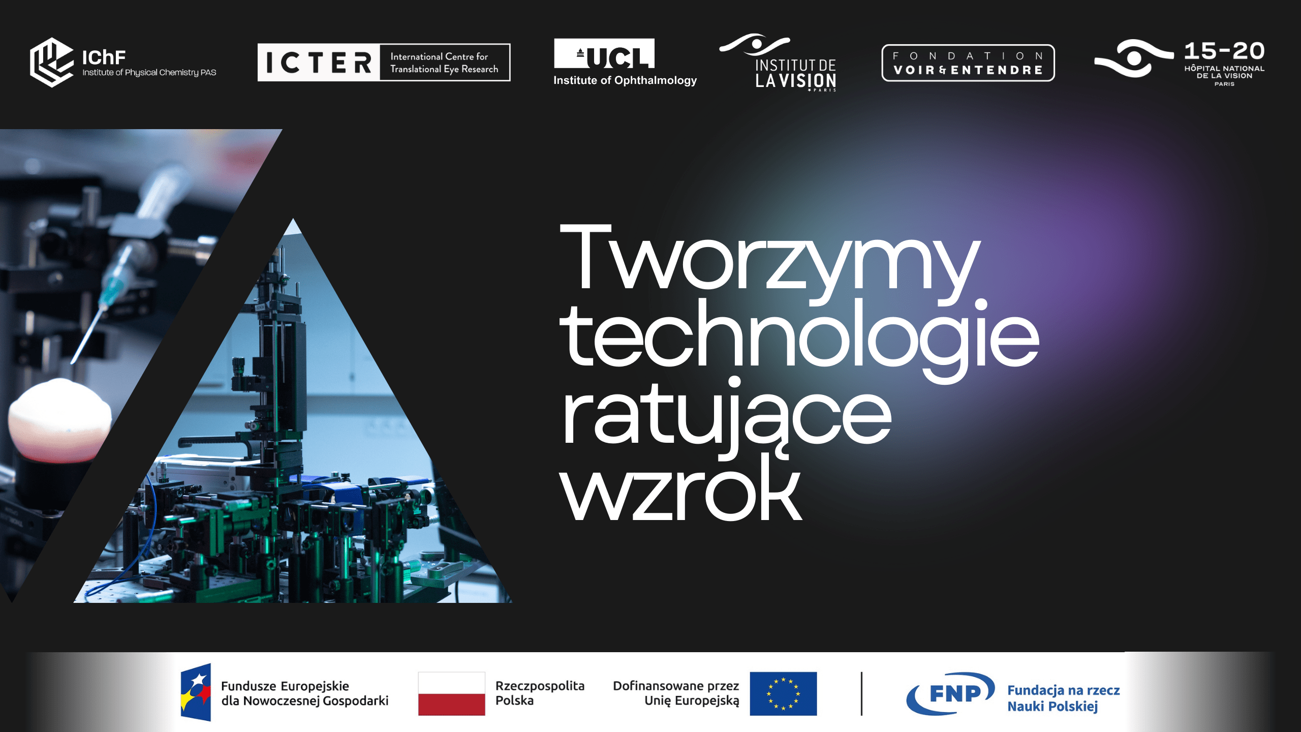
A special camera, laser and computer will show even more precisely what is happening inside our eyes - this is just one of the innovations from researchers at the ICTER science centre. We supported its team in applying for the prestigious 'Teaming of Excellence' grant. Now, the Warsaw scientists are creating an ophthalmic centre of scientific excellence together with researchers from Paris and London.
The goal of ICTER scientists is simple: to create solutions that will help ophthalmologists diagnose and treat patients' eyes. STOC-T technology, which will be able to replace today's OCT examination, commonly referred to as eye tomography, is now coming out of the laboratories into the world. The team that developed the new technology is headed by ICTER head Professor Maciej Wojtkowski. It was also he who created the first prototype of the OCT examination device 20 years ago, which is now used in eye clinics around the world.
The test itself is painless and takes 8.6 milliseconds to complete, which is the same as the double beat of the wings of a flying bee. The technology is expected to increase the detection of eye diseases at an early stage.
Capture the eye in stillness
- Timing is crucial in the examination of the eye. This organ constantly makes rapid movements that we are not aware of. Although we only needed a few seconds to carry out the examination with the current technology, each such movement introduced noise, which reduced the readability of the results, explains Professor Maciej Wojtkowski.
With help came the development of digital technology. The researchers used a camera that captures as many as 60,000 frames per second. This is more than a thousand times more than the standard cameras we use in our smartphones. This reduced the examination time to less than 0.01 seconds.
- In this fraction of a second, which is imperceptible to humans, the system we have created illuminates the eye with multiple waves of laser light. The camera captures hundreds of shots showing how the light propagates in the cells of the eye. The captured frames are then superimposed and analysed by our software, which creates a precise image of the eye's layers on this basis," says Professor Maciej Wojtkowski.
This will provide the ophthalmologist with an accurate image of the retina, on which he or she will be able to see signs of early development of many eye diseases, including glaucoma, macular degeneration or diabetic retinopathy. Increasing the accuracy of diagnosis is so important because today there are approximately 10,000 patients per ophthalmologist in Poland. According to the latest activity report of the National Health Fund, more than half a million Poles are waiting in line for an ophthalmology clinic. Meanwhile, making a diagnosis at an early stage of the disease will enable faster treatment, which in many cases can protect against loss of sight.
International support
The ICTER Science Centre is part of the Institute of Physical Chemistry of the Polish Academy of Sciences, and the scientists working there combine expertise in physics, biology, chemistry, engineering and medicine. It is this broad view of eye health problems that allows them to create new ways of investigating and treating vision.
The technology developed is one of the next steps planned by the researchers. Their main goal is to find a technological solution that will help to provide faster access to specialist ophthalmic care. Conducting such research was made possible by the prestigious grant they have just received in the 'Teaming for Excellence' programme in Horizon Europe. They have obtained funding of €30 million, of which €15 million is from the European Commission, with additional equivalent complementary funding from the Foundation for Polish Science and the Ministry of Science and Higher Education.
With this, ICTER will create a Centre of Scientific Excellence in Warsaw, a place where science meets practice and research is transformed into concrete medical solutions. Partners in this project are the Institute of Ophthalomogy at University College London and the Institut de la Vision at Sorbonne Université.

